Core Facility Session: Light Sheet
Friday, September, 30th, 2022: 11:00 am
Chairs: Alina Liebheit and Frank Schildberg
This year’s topic will spotlight the field of Light Sheet Fluorescence Microscopy (LSFM), a rapidly developing 3D imaging technique that is shared in core facility approaches around the globe. This technique provides a three-dimensional view into the inner working of the biological system. A prerequisite for this imaging method is a process called clearing, which renders the tissue transparent. Recent developments have produced a growing toolbox of tissue clearing, whole-mount immunostaining, and image analysis methods enabling the 3D exploration of almost every organ. The session will discuss how Light Sheet Fluorescence Microscopy can be used in human and mouse tissues when analyzing a biological question and will cover the strengths and limitations of the different methods.

From tissue clearing to cleared immunological processes
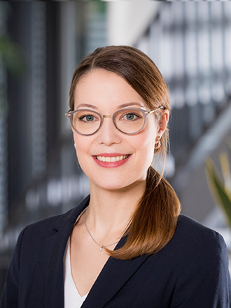
Leibniz-Institut für analytische Wissenschaften – ISAS – e.V., Dortmund
University Hospital Essen, University Duisburg-Essen, Germany
Abstract
Fluorescence microscopic analyses of large biological samples are subject to technical limitations due to their opacity, which causes light absorption, reflection and scattering. These physical characteristics are particularly challenging for analyses of anatomy and physiology. However, a new technology that overcomes these limitations is light-sheet fluorescence microscopy, which in combination with optical tissue clearing enables 3-dimensional analyses of large tissue samples. By using fluorescently labelled antibodies and obtaining endogenous fluorescence proteins, optical clearing methods together with light-sheet fluorescence microscopy (LSFM) enable the specific identification of biological structures down to individual cells.
The combination of optical clearing and LSFM represents a promising technology in basic and translational biomedical research. Based on a self-invented clearing protocol, we were able to identify and characterize for the first time a previously unexplained vascular system in long bones, which represents a significant contribution to the anatomical and physiological understanding of this organ. We were also able to identify a previously unknown macrophage population in knee joints that forms a physical barrier to the joint and shields it from inflammatory immune cells. This discovery opens up completely new therapeutic approaches for joint-associated diseases such as rheumatoid arthritis. These examples illustrate the wide range of applications of this technology and its high potential for future studies in the field of biology, translational research and clinical application.
Curriculum vitae
Academic Education and Degree
2020 Professor (W2), Leibniz-Institut für Analytische Wissenschaften – ISAS – e.V. Dortmund;
University Hospital Essen, University Duisburg-Essen, Germany
2017 Junior research group leader, Department of Internal Medicine 3, Rheumatology and
Immunology, UKER, FAU Erlangen-Nuremberg
2017 Postdoc, Department of Internal Medicine 3, Rheumatology and Immunology, UKER,
FAU Erlangen-Nuremberg
2017 Ph.D. thesis (Dr.rer.nat.), University Hosptial Essen, University Duisburg-Essen,
Germany
2011 Studies of Biology (M.Sc.), RWTH Aachen, Germany
Awards and Scholarships

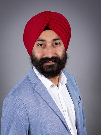
Spatial molecular profiling of 3D imaged whole organs and organisms
Institute for Tissue Engineering and Regenerative Medicine (iTERM), Helmholtz Zentrum München, Neuherberg, Germany.
Institute for Stroke and Dementia Research, Klinikum der Universität München, Ludwig-Maximilians University (LMU), Munich, Germany.
Abstract
While many diseases affect the entire body, research is usually conducted on the tissue of interest. To overcome potential bias, and discover previously unnoticed yet essential phenotypic changes in health and disease, we use a holistic approach. Towards this goal, we have developed novel tissue transparency methods- DISCO clearing, enabling in-depth antigen labeling, clearing and imaging of intact organs and organisms. DISCO clearing is fast (can be completed within a few hours) and versatile (applicable to various tissues including the brain, spinal cord, lung, heart, immune organs, and tumor bearing organs). We can now collect information at sub-cellular levels in whole organs and organisms without sectioning. Using DISCO clearing, we were able to characterize novel skull-dura connections and locate antibody-targeted vs. untargeted metastatic tumors in whole body of a mouse.
Moreover, to unravel the underlying molecular basis for the morphological changes in intact organs, we recently developed DISCO-MS. It’s a technology combining whole-organ/ism imaging, deep learning-based image analyses, micro tissue extraction and proteome analyses using ultra-high sensitivity mass spectrometry. DISCO-MS yielded qualitative and quantitative data indistinguishable from uncleared samples in both rodent and human tissues. We characterized microenvironment of early individual amyloid-beta plaques in Alzheimer’s disease mouse model. Furthermore, we studied regional proteome heterogeneity of immune cells in intact mouse bodies and aortic plaques in whole human heart. Thus, DISCO-MS enables unbiased proteome analysis of pre-clinical and clinical tissues after unbiased imaging of entire specimens in 3D, providing new diagnostic and therapeutic opportunities for complex diseases.
Short CV
2019-present: Omics Technologies Team Leader, Institute for Tissue Engineering & Regenerative Medicine (iTERM), Helmholtz Zentrum Munich, Neuherberg, Germany
2017-2019: Senior Researcher, Institute for Stroke and Dementia Research (ISD), LMU, Munich, Germany
2011-2017: Junior Group Leader, Neurochemistry Lab-1, University clinic Freiburg, Freiburg, Germany
2009-2011: Postdoctoral Fellow, University of California Los Angeles (UCLA), Los Angles, USA
2006-2009: Ph.D. Thesis (Dr.rer.nat.), Albert-Ludwigs University of Freiburg, Freiburg, Germany
2003-2005: Master’s in Biotechnology (M.Sc.), DIBNS Dehradun, HNBG University Srinagar, Garhwal, Uttarakhand, India

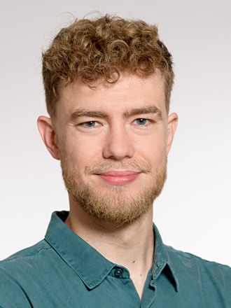
MarShie – a novel tissue clearing pipeline to comprehensively reveal myeloid-vasculature interactions throughout the murine bone marrow
Charité – Universitätsmedizin Berlin, Department of Rheumatology and Clinical Immunology, Berlin, Germany.
Abstract
Bone regeneration is a highly orchestrated process based on the micro-environmental interplay between the immune cell compartment, mesenchymal cells, and the vascular system. Around 5-10% of all fractures do not heal satisfactory and may result in mal- or nonunion. Refining our understanding of basic principles in the regeneration of bone potentially aids to the development of new regenerative strategies. Type H vessels, which are known to couple angiogenesis to osteogenesis during bone development are present in the fracture gap. The factors determining vessel growth under these conditions, as well as the complex spatial relationships between cellular compartments in the fracture gap have yet to be investigated in detail. We present here a novel tissue clearing pipeline termed MarShie (Marrow-Stabilization under harsh conditions via intramolecular epoxide linkage). This method enables us to analyze the three-dimensional interactions of myeloid cells with the vascular system, with cellular resolution. Our pipeline preserves endogenous fluorescence and diminishes light scattering and autofluorescence. Combined with light sheet fluorescence microscopy we used our pipeline to detect endogenous fluorescence of the myeloid compartment in CX3CR1-GFP+ cells, and the vascular network via Cdh5-tdTomato fluorescence. By using machine-learning based classifiers for cellular segmentation, we were able to quantitatively analyze these interactions in homeostatic state as well as in defect models for intramembranous and endochondral ossification. We observe that immediately after the tissue damage, myeloid cells sequester the area of bone injury. Later, they precede the growth of vessels and remain in close contact with endothelial cells throughout the regenerative process.
Biosketch
Till Mertens studied medicine at the Charité Berlin and the University of Córdoba. He started his medical doctorate in the group of Prof. Hauser in 2019, where he investigates the interactions between myeloid cells and the vasculature system during bone regeneration. Together with his colleague and PhD Student Alina Liebheit, he adapted a tissue clearing pipeline to render murine bones transparent. This novel method allows three-dimensional interrogation of the deep marrow at cellular resolution, unprecedented in deep bone marrow imaging.