European Guest Session: Austrian Society for Cytometry (OEGfZ)
Thursday, September, 29, 2022: 11:00 am
The Austrian Society for Cytometry offers a platform for everyone interested in flow cytometry and related fields such as fluorescence microscopy. The society sees itself as an interdisciplinary, integrative competence center in which information is developed, collected and passed on to the members in the most efficient way possible. This should promote a broader application of the knowledge and methods of this scientific area and guarantee an optimal use of the diverse possibilities. It is therefore a declared goal of the OEGfZ to build a bridge beyond the boundaries of existing specialist groups – between research, teaching and (routine) application.
The founding of the society in 2001 goes back to a series of activities that already took place in Austria in the 1990s. Ring-tests (inter-laboratory tests) have been carried out since 1992, the Vienna Flow Cytometry course (No Magic course) has been held since 1998, the magazine FlowMania appeared irregularly around 1999, and in April 2000 a regular cytometry meeting was initiated with the Flow Time event. Aware that there are several other dedicated cytometry users in Austria, the organizers of these events have decided to establish a society to create a common platform for such activities. The OEGfZ currently organizes cytometry congresses, leukemia-lymphoma workshops, cell culture days as well as basic and advanced courses in flow cytometry on a regular basis.
The Society’s goals should therefore appeal to all those working in the field of cellular characterization. This includes students as well as representatives of technical professions, natural scientists and physicians of all disciplines.
In such a technology-intensive area, in which the availability and quality of the appropriate devices and reagents play an essential role in successful work, close cooperation with the manufacturers and producers is of great importance. The OEGfZ understands the exchange with the companies, some of which are the main sponsors of the society, as a very fruitful cooperation on a very friendly basis.

Introduction: The Austrian Society for Cytometry
Core Facility Flow Cytometry & Surgical Research Laboratories, Medical University of Vienna, Austr
Biosketch
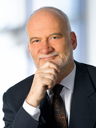
Andreas Spittler received his diploma in medicine and his habilitation in Pathophysiology at the Medical University of Vienna. He started his research career in 1991 at the Department of Pathophysiology and was appointed Associate Scientist at the Surgical Research Laboratories in 1998. At that time, he investigated the influence of amino acids on monocyte immune regulation in both cell culture and patient-oriented studies. Since 2008 he has headed the Core Facility Flow Cytometry. Andreas Spittler was a founding member of the Austrian Society for Cytometry (current President) and of the Austrian Society for Extracellular Vesicles. His main scientific interests are in the characterization of extracellular vesicles using flow cytometry and imaging flow cytometry, as well as in the characterization of the neonatal immune system and the functional characterization of monocytes in inflammation and sepsis.

One tube to find them all
Internal Medicine V, Hematology and Oncology, Medical University Innsbruck and Tyrolean Cancer Research Institute, Innsbruck, Austria
Abstract
High-dimensional single cell analysis has provided unprecedented insights into the cellular makeup of heterogeneous samples. However, especially in clinical and translational applications, where robust assessment of a high number of samples even in multicenter settings is necessary, information content and practicability have to be carefully weighted.
Using a carefully selected combination of 14 surface markers, we designed a multicolor panel to identify and quantify all principal leukocyte populations, compatible with standard flow cytometric instruments and thus accessible to a particularly large research community. Optimized for use in whole blood, this panel is well-suited for identification and absolute cell counting of neutrophils, eosinophils, basophils, T cells, natural killer cells, B cells, plasma cells, monocytes, myeloid and plasmacytoid dendritic cell populations as well as progenitor cells. Separation of populations is high and virtually no cells remain undefined after gating. In addition, promiscuous expression of some antigens allows determination of subpopulations and activation status.
For the analysis of tissue derived cells, we have expanded the panel to include live/dead discriminators and markers for epithelial, endothelial and mesenchymal cells. Using this panel for orthogonal validation of single cell sequencing data, we demonstrate that not all cell populations are captured equally well by different platforms.
In summary, we present a versatile panel for immune profiling on broadly available instrumentation, which can also be used as a backbone for more sophisticated investigations.
Biosketch
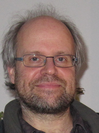
Currently, Sieghart Sopper is head of the Flow Cytometry Unit and coordinator of the research laboratories of the Clinic for Hematology and Oncology at the Medical University Innsbruck. He is also affiliated as a group leader to the Tyrolean Cancer Research Institute. He studied Biology at the University of Graz and did his doctoral thesis at the University of Würzburg, where he also habilitated in the field of Virology. He worked as independent group leader at the Universities of Würzburg and Göttingen as well as at the DPZ – the Leibniz Institute for Primate Research. Using various single cell technologies, he investigates the role of the immune system in infectious diseases and cancer in several preclinical and translational studies.

Thinking outside the box in cell culture
Core Facility Alternative Biomodels & Preclinical Imaging, Medical University of Graz, Graz, Austria
Abstract
Sarcomas originate from a diversity of mesenchymal tissue lineages including muscle, cartilage, bone or fibrous and can therefore occur in almost all organs. Most common histological subtypes include liposarcoma, chondrosarcoma, osteosarcoma and Ewing sarcoma, with the latter two being primarily pediatric tumors. A major obstacle to drug development and subsequent preclinical studies is the lack of suitable in vitro models due to the rare incidence of each sarcoma subtype. In this study post-surgical tumor tissue from a multitude of sarcoma entities was enzymatically and mechanically dissociated. In addition to the tumor tissue, surrounding tumor tissue and/or healthy skin tissue was collected and immortalized. The obtained patient-derived primary cells underwent extensive quality control and detailed characterization by using various technologies. Specifically, we would like to present the clear cell sarcoma lines MUG Lucifer prim and MUG Lucifer met, which allow translational research in terms of innovative treatment strategies and the study of the metastatic process. Particularly exciting is the release of extracellular vesicles from the established sarcoma cell lines, where we have been able to achieve robust reproducible results using the ExoView technology. With recent advances in the field of cancer research, the use of complex patient derived models has become increasingly popular and important. The biology of tumor diseases, the communication of tumor cells with their environment, tumor progression and especially metastasis behavior can only be successfully studied and captured with adequate models.
Biosketch
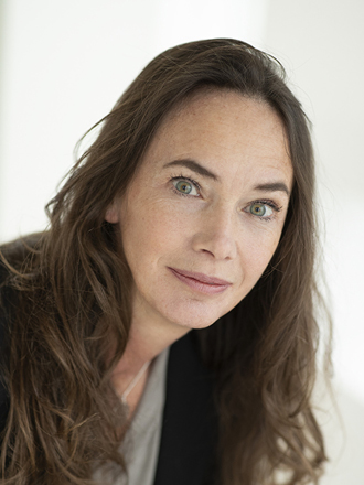
Beate Rinner is head of the core facility Alternative Biomodels and Preclinical Imaging (CF ABPCI) at the Division of Biomedical Research. Her group focuses on the establishment and characterization of rare tumor cell lines. Furthermore, the isolation of cancer associated fibroblasts (CAFs) and immortalization by using hTERT to create autologous cancer models for drug screening and tumor biology investigation. She focuses on the communication between tumor cells and CAFs, by isolation and detail characterization (using NanoSight, flow cytometry, westernblot, qPCR and NanoView technology) of extracellular vesicles and supernatant investigation by using xMAP technology. She has extensive experience on cell line establishment, tumorigenicity testing and pharmacological efficacy studies furthermore she uses the most modern techniques, (micro-ultrasound, micro-computed tomography and optical imaging) to obtain images on a wide variety of levels from full-body imaging to subcellular structures.

Mapping the cellular landscape of human airways by using mass cytometry
Core Facility Flow Cytometry, Medical University of Vienna, Austria
Biosketch
Lena Müller obtained her PhD from the Medical University of Vienna, Austria. Her research was on understanding the molecular mechanisms of T cell development under the supervision of Prof. Willfried Ellmeier (Institute of Immunology). She then joined the laboratory of Dr. Michael Bonelli (Department of Rheumatology/ General Hospital of Vienna) to investigate T cells in the context of autoimmunity. During her research projects she gained an extensive expertise in assay development focusing on phenotyping of immune cells. In 2021 she joined the Core Facilities at the Medical University of Vienna and started to work with mass cytometry. She is coordinating the application of mass cytometry on various projects and operates the Helios/ Hyperion instrument.