Imaging Session:
Wednesday, September, 28th, 2022: 12:00 pm
Chairs: Raluca Niesner and Anja Hauser
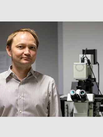
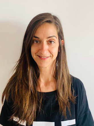
In line with the theme of the conference “Spotlight on cells-lifestyle and environment”, the imaging session is focusing on various ways to analyze the interaction of cells with their microenvironment and cellular responses to various stimuli coming from their surroundings. The session will cover a variety of experimental imaging systems, ranging from single cells in microfluidic systems via tissue sections and organoids, to intravital microscopy. We will start with a talk by Prof. Dmitriev, who is using fluorescence and phosphorescence lifetime imaging to analyze tissue oxygenation and metabolism. Our second speaker is Dr. Anna Pascual-Reguant who is combining various spatial technologies, at the protein and transcriptomic level, to analyze COVID-19 lung pathology. Alexander Fiedler will tell us about a fluorescence lifetime (FLIM) based metabolic analyses in vivo, which he longitudinally performs in the context of bone regeneration. Our fourth speaker, Eric Sündermann, is reporting on the possibility to use FLIM to analyze the mechanical properties of cells. We are looking forward to an exciting session about cytometry in context.

Probing intestinal organoids oxygenation and metabolism using phosphorescence and fluorescence lifetime imaging microscopies
Tissue Engineering and Biomaterials Research Group, Dept. Human Structure and Repair, Ghent University, Ghent, Belgium
Abstract
Success in studies of 3D (micro)tissues and organoids is often hampered by their intrinsic heterogeneity and dynamic gradients of biomolecules. Dynamics of cell metabolism, excretion products and O2 are hard to predict and are typically controlled by western blotting, next generation genome, RNA sequencing and traditional antibody-based microscopy methods. My team addresses the challenge of non-destructive quantitative multi-parametric imaging of 3D tissue models by using high-performance nanoparticles, new sensor chemistries and live fluorescence (FLIM) and phosphorescence (PLIM) lifetime imaging microscopies. In my talk I will briefly introduce the methodology of O2 sensing by PLIM and how this methodology can help visualizing live oxygenation of mouse adult stem cell derived intestinal organoids. Using this approach, complemented by the two-photon excited NAD(P)H autofluorescence imaging and other FLIM methods, we found that stem cell metabolism strongly depends on the nutrient availability in the growth medium. Collectively, presented methodology is highly useful for studies of the stem cell niche metabolism in the organoid cultures.
Biosketch
Ruslan I. Dmitriev is an Assistant Professor in the Department of Human Structure and Repair, Ghent University, Ghent, Belgium. He received his Ph.D. in bioorganic chemistry from the Shemyakin-Ovchinnikov Institute of Bioorganic Chemistry, Russian Academy of Sciences, and continued his postdoctoral training at the University College Cork, Ireland. Before joining Ghent University in 2020, Ruslan was a staring investigator-research fellow at the Metabolic Imaging Group, University College Cork. Dr. Dmitriev’s research interests are in imaging of cellular oxygen gradients and cell metabolism in the three-dimensional tissue models, such as tumor and stem cell-derived spheroids and intestinal organoids, using multi-parameter live cell fluorescence lifetime imaging microscopy (FLIM) and high-performance protein-, bioconjugate- and nanoparticle-based biosensors. Dr. Dmitriev current work aims at developing advanced tools to study cell metabolic requirements in the tumor and stem cell-derived spheroids, intestinal organoids and addressing their heterogeneity using imaging-assisted tissue engineering tools.

Activated fibrovascular niches orchestrate immunopathology in severe COVID-19 disease
AG Hauser, Immune Dynamics, Deutsches Rheuma-Forschungszentrum & Charité-Universitätsmedizin Berlin
Abstract
Post-acute lung sequelae of COVID-19 are challenging many survivors across the world, yet the mechanisms behind are poorly understood. We have analyzed post-mortem lung and lymph node tissue of COVID-19 cases and non-COVID-related pneumonia controls, by using several state-of-the-art imaging techniques, snRNAseq and spatial transcriptomics. We have stratified the COVID-19 donors based on disease duration and included a prolonged group (7 – 15 weeks of disease duration post-infection), where viral infection had been resolved. Nevertheless, prolonged COVID-19 lungs showed persistent tissue damage along with an increase in lymphocyte infiltration, features that recapitulate those previously reported for Long-COVID.
Our unique spatial approaches reveal that, upon endothelial dysfunction, activated fibrovascular niches emerge as crucial sites, where the molecular mechanisms driving a dysregulated interaction between immune cells and the surrounding tissue occur. We pinpoint two main factors, CCL18 and CCL21, which modulate the local stromal composition and favor endothelial to mesenchymal transition, resulting in the expansion of fibrovascular niches. This, in turn, results in an excessive accumulation of CCR7+ T cells, which become locally imprinted with an exhausted, T follicular helper-like phenotype, as opposed to the sustained T cell activation found in draining lymph nodes. The perpetuation of local chronic inflammation, which is independent of viral persistence, results in the formation of tertiary lymphoid structures as the ultimate manifestation of tissue repurposing.
Biosketch
I am a postdoctoral researcher in the Hauser Lab (Immune Dynamics) at the Deutsches Rheuma-Forschungszentrum and the Charité-Universitätsmedizin Berlin. I received my PhD from the Department of Biology, Chemistry and Pharmacy of Freie Universität Berlin. My research focuses on the in-depth and spatially resolved analysis of local immune responses using state-of-the-art, multiplexing techniques. I am interested in studying tissue niches, both at the protein and transcriptional level, as self-organized functional units that modulate local immunity and dictate tissue homeostasis and disease. I am particularly keen on sharing my enthusiasm for spatial immunology and microscopy with my colleagues and working in interdisciplinary environments.
Alexander Fiedler (Short Talk)
Deutsches Rheuma-Forschungszentrum Berlin, ein Leibniz-Institut, Biophysical Analytics, Berlin, Germany
Development of functional in vivo fluorescence lifetime microendoscopy of the femoral bone marrow
Immune cell (dys)function is closely related to their metabolism. However, the particular cause and mechanisms remain elusive, mainly due to the lack of technologies to visualize local metabolism in vivo over long periods.
My project is about the development of Fluorescence-lifetime Longitudinal Intravital Microendoscopy of the Bone marrow (FLIMB). This novel method will combine fluorescence lifetime imaging and longitudinal micro-endoscopy of the bone marrow (LIMB) in live mice: Multi-photon microscopy and implant-based endoscopic gradient optics allow very deep and dynamic fluorescence imaging of the bone marrow tissue and the finely orchestrated self-organization of (immune) cells (e.g. re-vascularization after injury). Additionally, the fluorescence lifetime imaging of the autofluorescent metabolic molecules NADH and NADPH will allow the simultaneous analysis of the local cellular (immuno)metabolism and function, respectively.
With FLIMB, we found dynamic metabolic and functional heterogeneity of myeloid cells during bone regeneration after osteotomy. We further aim to gain deeper insights into spatiotemporal and functional aspects of bone marrow biology during health and disease and expect that environmental factors (like nutrient and oxygen availability and the inflammatory milieu) determine metabolic activity, which impacts on the diverse functions myeloid cells fulfil.
Eric Sündermann (Short Talk)
ZIK HIKE, Institute of Physics, University of Greifswald, Greifswald, Germany
Cytometric single-cell analysis of membrane tension
Real-time deformability cytometry (RT-DC) is a biomechanical method which is able to characterise the physical properties of cells. To do so, cells travel through a microfluidic chip assembled on an inverted microscope. Every cell is imaged by a high-speed camera, and a convex contour is fitted to its shape to calculate the corresponding deformation. While mechanical properties of cells can be derived from analytical models and knowing the deformation as well as the hydrodynamic stress distribution, this approach is not suitable to discriminate between membrane and cytoskeletal contributions toward cell mechanics.
Here, we aim to shed light on dynamic alterations in membrane tension of cells passing a microfluidic constriction by introducing a new method that combines cytometry with fluorescence lifetime imaging microscopy (FLIM). As a fluorescent probe we applied Flipper-TR that is incorporated in the lipid-bilayer membrane of cells. In preliminary experiments, we measured the membrane tension of myeloid precursor cells (HL60 cells) at various hydrodynamic and osmotic stresses. We are able to observe a stress-dependent membrane tension response. We also found a change in the membrane tension for dimethyl sulfoxide (DMSO), which influences the membrane fluidity, but not for the exposure with cytochalasin D, which alters the level of polymerization of the underlying actin cortex. In future studies, we want to complement FLIM with RT-DC to simultaneously measure the contribution of membrane tension and the cytoskeleton to the mechanical properties of cells.